Minggu, 28 Oktober 2012

 PAVING BREAKER B40


KAMI PT. FAJARMASMURNI AGEN RESMI (TUNGGAL) AIRMAN JAPAN WILAYAH INDONESIA KALAU BERMINAT DAN INGIN TAU TENTANG AIRMAN HUBUNG SAYA 
AGUSTIAR-PRODUCT ENGINEER
CP 085260025094 atau
EMAIL :  agustiar@fajarmasmurni.com
atau kunjungi WWW.FAJARMASMURNI.COM

Rabu, 24 Oktober 2012

FURUKAWA PCR200

 FURUKAWA ROCK DRILL PCR200


KAMI PT. FAJARMASMURNI AGEN RESMI (TUNGGAL) AIRMAN JAPAN WILAYAH INDONESIA KALAU BERMINAT DAN INGIN TAU TENTANG AIRMAN HUBUNG SAYA 
AGUSTIAR-PRODUCT ENGINEER
CP 085260025094 atau
EMAIL :  agustiar@fajarmasmurni.com
atau kunjungi WWW.FAJARMASMURNI.COM

Generator AIRMAN

 GENERATOR SDG100AS 


KAMI PT. FAJARMASMURNI AGEN RESMI (TUNGGAL) AIRMAN JAPAN WILAYAH INDONESIA KALAU BERMINAT DAN INGIN TAU TENTANG AIRMAN HUBUNG SAYA 
AGUSTIAR-PRODUCT ENGINEER
CP 085260025094 atau
EMAIL :  agustiar@fajarmasmurni.com
atau kunjungi WWW.FAJARMASMURNI.COM

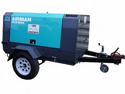
Compressor AIRMAN

Compressor AIRMAN JAPAN PDS185S


KAMI PT. FAJARMASMURNI AGEN RESMI (TUNGGAL) AIRMAN JAPAN WILAYAH INDONESIA KALAU BERMINAT DAN INGIN TAU TENTANG AIRMAN HUBUNG SAYA 
AGUSTIAR-PRODUCT ENGINEER
CP 085260025094 atau
EMAIL :  agustiar@fajarmasmurni.com
atau kunjungi WWW.FAJARMASMURNI.COM

Mikroskop VIVAVIEW FL INCUBATOR FLUORESCENCe


VIVAVIEW FL INCUBATOR FLUORESCENCE MICROSCOPE OVERVIEW

  • Ideal time-lapse imaging inside an incubator
  • Parallel time-lapse observation of eight dishes
  • Eliminates thermal drift during long-term imaging
  • High quality fluorescence and DIC observation
  • Cooled CCD camera
  • Stress-free time-lapse imaging
  • Simple computer based control
  • Powerful and proven image analysis with MetaMorph for Olympus
  • Ability to load chemical solutions during growth

VIVAVIEW FL INCUBATOR FLOURESCENCE MICROSCOPE SPECIFICATIONS

Objective UPLSAPO40x (NA 0.95, WD 0.18mm)
Intermediate magnifications 0.5x/1x/2x, Motorized magnification changer
Morphological observation (Transmitted light) Light source 1W LED (625nm)
Contrast method Differential Interference Contrast (DIC)
Condenser NA 0.55, FS fixed (rectangle), DIC prism fixed
Fluorescence Observation (incident light) Light Source X-Cite eXacte Pre-aligned 2000 hour DC lamp with Closed - Loop Feedback, Adjustable Iris, Liquid Light Guide
Excitation change Motorized 6 position, 1 for transmitted light
Motorized Stage Stroke 10mm x10mm, each dish
Sample Capacity Glass bottom dishes with 35mm diameter, up to 8
Sample Tray Can be easily put on and taken off, autoclave capability, include 8 lids with glass window for the glass bottom dishes with 35mm diameter.
Focus
Objective drive, motorized up/down control
Operation Jog dial control
Camera Cooled CCD camera, -35oC air cooled
2/3" (8.67mm x 6.60mm), 1344 x 1024 pixels
12 bits or 16 bits
Liquid handling assist (BTO option) Motorized open/close control of designated dish. Manual pipetting through the small opening on the internal door of the incubator.
Imaging software MetaMorph
CO2Incubator Temperature 37°C
Accuracy +/- 0.3°C (ambient temperature 25oC, no load)
Humidity 95% +/- 5% RH
CO2Concentration 0-20%
Accuracy +/- 0.15% (ambient temperature 25°C, no load)
Operating conditions Temperature 15°-28°C
Humidity Less than 80% RH
Dimensions of main body 710mm x 630mm x 1170mm
Weight 150kg
Electricity power consumption Main body 500VA
PC and monitor About 400VA


Aplikasinya

VIVAVIEW FL INCUBATOR FLUORESCENCE MICROSCOPE APPLICATIONS

Click Here to view VivaView FL Video Gallery

PUBLICATIONS

  • Meeting the Challenges of Long-term Time-lapse Imaging - Bioscience Technology Sept 2009 (PDF) Read More >
  • Takahiro Seki, Takayuki Shimahara, Kazuhiro Yamamoto, Nana Abe, Taku Amano, Naoko Adachi, Hideyuki Takahashi, Kaori Kashiwagi, Naoaki Saito, Norio Sakai. (2009). Mutant ?PKC found in spinocerebellar ataxia type 14 induces aggregate-independent maldevelopment of dendrites in primary cultured Purkinje cells. Neurobiology of Disease. Vol. 33, 260-273. Read More>
  • Asako Sakaue-Sawano, Hiroshi Kurokawa, Toshifumi Morimura, Aki Hanyu, Hiroshi Hama, Hatsuki Osawa, Saori Kashiwagi, Kiyoko Fukami, Takaki Miyata, Hiroyuki Miyoshi, Takeshi Imamura, Masaharu Ogawa, Hisao Masai, and Atsushi Miyawaki. (2008). Visualizing Spatiotemporal Dynamics of Multicellular Cell-Cycle Progression. Cell. Vol. 132, 487-498. Read More>
  • Asako Sakaue-Sawano, Kenji Ohtawa, Hiroshi Hama, Masako Kawano, Masaharu Ogawa, and Atsushi Miyawaki. (2008). Tracing the Silhouette of Individual Cells in S/G2/M Phases with Fluorescence. Chemistry & Biology. Vol. 15, 1243-1248. Read More>
  • Hiroki Umeshima, Tomoo Hirano, and Mineko Kengaku (2007). Microtubule-based nuclear movement occurs independently of centrosome positioning in migrating neurons.
    PNAS. Vol.104, No. 41, 16182-16187. Read More>
  • Takahiro Hatanaka, Yasue Hatanaka, Jun-ichi Tsuchida, Vadivel Ganapathy, and Mitsutoshi Setou. (2006) Amino Acid Transporter ATA2 Is Stored at the trans-Golgi Network and Released by Insulin Stimulus in Adipocytes. Journal of Biological Chemistry. Vol. 281, No. 51, 39273-39284. Read More>

VIDEOS

Screening drugs to block aggregation of Huntingtin protein

Apoptosis: An award-winning video

Instrument: VivaView FL (LCV-110)
Subject: This video is a series of live, time-lapse images of human mesenchymal stem cells grown on a collagen-I coated dish. The cells are treated with a high level of a PI3-Kinase inhibitor which induces apoptosis (or programmed cell death). This video won an award at the 2010 American Society for Cell Biology Cell Dance competition.
Reference: Video courtesy of Ms. Kira Henderson, PhD student, Department of Biology, Rennselaer Polytechnic Institute, Troy, NY.
PLAY VIDEO
Screening drugs to block aggregation of Huntingtin protein

Screening drugs to block aggregation of Huntingtin protein

Instrument: VivaView FL (LCV-110)
Subject: Montage displaying aggregate accumulation of GFP-tagged truncated Huntingtin in PC12 cells under 3 different concentrations of an aggregation inhibitor drug vs. control media.
Reference: Emily Mitchell, Leslie Thompson, Ph.D., University of California, Irvine
PLAY VIDEO
Human neural stem cells are healthy during two weeks of imaging

Human neural stem cells are healthy during two weeks of imaging

Instrument: VivaView FL (LCV-110)
Subject: 2 week time lapse of Human Neural Stem cells transfected with GFP CAG. Development is followed after the addition of differentiation media at T = 24 hours.
Reference: Chris Sontag, Brian J. Cummings, Ph.D., University of California, Irvine
PLAY VIDEO
Comparing effects of immunosuppressant drugs on human neural stem cell fate

Comparing effects of immunosuppressant drugs on human neural stem cell fate

Instrument: VivaView FL (LCV-110)
Subject: Immunosuppressant drug study of Human Neural Stem cells to analyze proliferation and migration. Movie shows a 2 x 2 montage from a single specimen dish. Focus is maintained throughout as new drug and media is added every 3 days.
Reference: Chris Sontag, Brian J. Cummings, Ph.D., University of California, Irvine
PLAY VIDEO
Migration tracking of human neural stem cells

Migration tracking of human neural stem cells

Instrument: VivaView FL (LCV-110)
Subject: Tracking data of Human Neural Stem cells captured at 15 frames per second.
Reference: Chris Sontag, Brian J. Cummings, Ph.D., University of California, Irvine
PLAY VIDEO

Biocompare Video for VivaView FL Incubator Fluorescence Microscope

PLAY VIDEO

Hela Cells constantly-expressing Fucci

Instrument: VivaView FL (LCV-110)
Subject: VivaView FL makes a contribution as the microscope to provide a stable environment in culture for the long term. 64 hours 13.5 minutes.
Reference: Sakaue-Sawano A, Atsushi Miyawaki M.D., Ph.D. RIKEN Brain Science Institute, Laboratory for Cell Function Dynamics
PLAY VIDEO

HeLa Cells expressing Histone H2B-GFP Cells divide 3 times during 60 hours of fluorescence and DIC observation

Instrument: VivaView FL (LCV-110)
Subject: Successful observation of 3 divisions in cells expressing GFP localized to the nucleus is challenging for fluorescence microscopy. The VivaView FL allows long-term imaging by minimizing damage to the cells while maintaining image qality.
Reference: Atsushi Miyawaki M.D., Ph.D. RIKEN Brain Science Institute, Laboratory for Cell Function Dynamics
PLAY VIDEO

Purkinje neuron

Instrument: VivaView FL (LCV-110)
Subject: Fluorescence time lapse observation of primary cell cultures is extremely difficult. Using the VivaView FL, Dr. Kengaku successfully captures a 4 days time-lapse data series of a neuron.
Reference: Mineko Kengaku Ph.D., RIKEN Brain Science Institute, Laboratory for Neural Cell Polarity
PLAY VIDEO

Superior cervical ganglion cells

Instrument: VivaView FL (LCV-110)
Subject: Transportation of Mitochondria in neurites. The VivaView FL captures mitochondrial movement that is faster than previous reported.
Reference: Mitsutoshi Setou, M.D., Ph.D., Molecular Gerontology, Mitsubishi Kagaku Institute of Life Sciences.
PLAY VIDEO

PC12 cell with GFP - Model system for neuronal differentiation

Instrument: VivaView FL (LCV-110)
Subject: 6 daytime-lapse data captured at 2 hour intervals. The VivaView FL maintains cell viability for over 6 days of imaging.
PLAY VIDEO

KAMI PT. FAJARMASMURNI AGEN RESMI (TUNGGAL) MIKROSKOP OLYMPUS JAPAN WILAYAH INDONESIA KALAU BERMINAT DAN INGIN TAU TENTANG OLYMPUS HUBUNG SAYA 
AGUSTIAR-PRODUCT ENGINEER
CP 085260025094 atau
EMAIL :  agustiar@fajarmasmurni.com
atau kunjungi WWW.FAJARMASMURNI.COM

 Kamera DP 21

DP21® DIGITAL CAMERA OVERVIEW

Highlights

  • 2 Megapixel easy-to-use, stand-alone camera system; records images to USB Drive.
  • 12-bit color depth for outstanding color fidelity
  • 15 frames per second live image preview at full resolution (1600 x 1200 UXGA)
  • Color or monochrome mode
  • IEEE 1394a connectivity with optional software
  • Automatic exposure and white balance. Manual mode for fine tuning
  • 2X and 4X live zoom preview with panning
  • 5 image quality modes: 1600 x 1200 SHQ TIFF or JPEG, 1600 x 1200 HQ JPEG, and 1280 x 960 SQ JPEG (high and low compression)
  • 3 selectable ISO sensitivities; 100, 200, and 400
  • Exposure times ranging from 1/20,000 sec. to 2 sec.
  • Autofocus aid for easy focusing
  • Selectable scale bar settings
  • Twelve types of measurement tools
  • Video recording capability of up to 5 minutes

Color fidelity

The 2 megapixel DP21 uses a 1200 x 1600 pixel Bayer mask CCD and interfaces to a computer via IEEE 1394a technology for rapid image download. It uses a 12-bit analog to digital converter, recording subtle color differences important for accurate color reproduction. The result is images with outstanding color and fine detail.

Flexibility

Designed with the same photomicrography concept as other popular Olympus cameras, the DP21 is offered as an easy-to-use, stand-alone solution; ideal when precious bench space is limited. The camera is controlled from a convenient stand-alone handset control box which is newly designed and ergonomic; operation is smooth with the functional key layout. Imaging is quick and easy. Set the camera to automatically expose and white balance the image or fine tune the exposure manually. An over/under exposure notification ensures images are perfect every time.

Software control

The DP21 digital camera can be controlled by Olympus cellSens® Digital Imaging Software.

DP21 Graphical User Interface

The DP21 digital camera offers an assortment of simple tools that are designed to streamline workflow. A focus indicator combined with the 2X and 4X digital zoom and rapid display make framing and focusing effortless. A scale bar may be overlayed on the image with settings for up to seven magnifications. There are twelve simple measurement tools.

CKX41 & CKX31 Mikroskop


Gambar CKX31


Gambar CKX41


CKX41 & CKX31 MICROSCOPE OVERVIEW

  • CKX31 & CKX41 are designed using the (UIS2) infinity-corrected optical system
  • A robust, small footprint frame with triangular base and solid front-to-back support provides exceptional stability and allows the user more room on the tabletop
  • Improved optical performance for Phase Contrast and new Relief Contrast
  • A pre-centered slider saves time in configuration of the phase annulus
  • Unique tilting observation tube offers ergonomic positioning for sitting and standing observation (optional on the CKX41 only)
  • Powerful 6V/30W halogen bulb provides significantly enhanced image quality and brightness for observation of cells
  • Glass stage insert provides quick recognition of objective lenses (optional on CKX41 only)
  • Features a fluorescence illuminator; a dual slider for blue and green excitation cubes, and offers a cost effective 50w mercury system (optional on the CKX41 only)
  • Centering-free, common phase annulus for 10x, 20x, and 40x; needs no adjustment (standard with the CKX31)

CKX41 & CKX31 Microscope Specifications

Item CKX41 CKX31
Optical system UIS2 (Universal Infinity-corrected System) optics
Focus Vertical nosepiece movement (stage is fixed), coaxial coarse and fine focus with tension adjustment mechanism, roller slide mechanism, stroke: 7mm up and 2mm down from focus position which is 1mm above the stage, stroke per rotation: 39.6mm (coarse), 0.2mm (fine)
Revolving nosepiece Quadruple
Stage Plain Stage 160mm(L) x 250mm(W), stage clips attachable
Exchangeable insert plate (Ø25mm opening) incorporated
Mechanical stage Right-hand low drive coaxial controls, stage movement: X=120mm, Y=78mm, with three dish/sample holders
Substage 70mm(L) x 180mm(W)
Illumination system Light source 6V/30W halogen lamp, lamp socket (U-LS30-3-2), built-in frosted and heat absorbing filters, detachable illuminator
Filter holder Insert up to 11mm thick with Ø45mm filter, detachable
Aperture diaphragm Lever type, range: from minimum Ø3mm to maximum Ø44mm
Slider insertion With phase slider pocket and built-in slider position click stop mechanism
Condenser Detachable ultra-long working distance condenser (N.A. 0.3, W.D. 72mm)
Contrast slider Pre-centered Phase Contrast: 4x, 10x/20x/40x, empty slot Centerable Phase Contrast: 4x, 10x/20x, empty slot (40x optional, pre-centered)
Observation tube Binocular tube(U-CBI30-2) Inclined 30°, interpupillary distance range: 48mm-75mm, diopter adjustment by helicoid on left sleeve Fixed binocular tube, Inclined 45°, interpupillary distance range: 48mm-75mm, diopter adjustment by helicoid on right sleeve
Trinocular tube(U-CTR30-2) Inclined 30º, ring dovetail attachment, interpupillary distance range: 48-75mm, tube length and diopter adjustment by helicoid on left sleeve. Observation optical path: 50 (binocular)/50 (video port)
Tilting binocular Tube(U-TBI-3) Variable inclination angles from 5º to 35º, circular mounting dovetail attachment, interpupillary distance range: 50mm-76mm, diopter adjustment by helicoid on right sleeve.
Vertical fluorescence illuminator Detachable illuminator, switchable slide (2 position: Blue excitation, Green excitation, transmitter light observations. Two cubes attachable simultaneously), built-in UV absorbing filter
Eyepiece WHB10X/WHB10X-H (F.N. 20) for U-CBI30 and U-CTR30WHB10X/WH10X-H/35WH10X/PWH10X (F.N. 22) for U-TBI-3 10X (F.N. 20)
Power supply Continuous intensity adjustment, built-in voltage changeover switch (100/120V, 220/240V), frequency 50/60Hz


CKX41 ACCESSORIES

  • Ergonomic - The optional tilting binocular tube reduces neck strain, and low positioning of the stage handle and focus knob, and frontal light control make long term use comfortable.
  • Documentation and analysis - Olympus digital cameras can be adapted via the optional trinocular observation tubes. Choose the Olympus camera model which fits best your needs.
  • Cost effective fluorescence - Fluorescence labeling methods are more frequently used in routine observations. Olympus offers a cost-effective 50 Watt Mercury illumination system suitable with dual filter slider for fluorescence and GFP observations.
  • Easy contrast - Speed up your observation with the pre-centered phase contrast. Visualize your specimen under different magnifications without time-consuming adjustments. Increase visibility and contrast in living and unstained material particularly if plastic cell culture devices are used. Alternatively, the relief contrast technique creates apparent three-dimensional images.
  • Documentation and analysis - Olympus digital cameras can be added via the optional trinocular observation tubes. Add one of our software packages for capture, analysis and reporting of your data.

CKX31 ACCESSORIES

  • Simplicity - Standard microscope for cell culture observation with fixed binocular tube.
  • Easy contrast - Speed up your observation with the pre-centered phase contrast. Visualize your specimen under different magnifications without time-consuming adjustments. Increase visibility and contrast in living and unstained material particularly if plastic cell culture devices are used. Alternatively, the relief contrast technique creates apparent three-dimensional images.

Peripherals

Optics

The new UIS2 PlanCN Series of objectives replaces the UIS Achromat objectives as the standard for this microscope. Benefit from the plan correction over the complete field of view for efficient observation and documentation.

KAMI PT. FAJARMASMURNI AGEN RESMI (TUNGGAL) MIKROSKOP OLYMPUS JAPAN WILAYAH INDONESIA KALAU BERMINAT DAN INGIN TAU TENTANG OLYMPUS HUBUNG SAYA 
AGUSTIAR-PRODUCT ENGINEER
CP 085260025094 atau
EMAIL :  agustiar@fajarmasmurni.com
atau kunjungi WWW.FAJARMASMURNI.COM